Brain Ventricle Enlargement Suggests Which of the Following
The ventricular volume including lateral 3rd and 4th ventricles showed a remarkable difference among HCs MS and NMOSD patients. 2002 first-episode schizophrenia Fannon et al.

Conventional Diagnostic Coronary Angiogram And Coronary Ct Angiogram Cardiac Event Cardiac Catheterization Cardiac Cycle
Enlarged ventricles in the brain may be a sign of normal pressure hydrocephalus.

. The third ventricle. The diagnosis of atrophy of the brain based on the visual interpretation of CT findings appears questionable. The fourth ventricle.
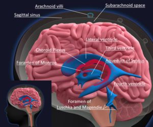
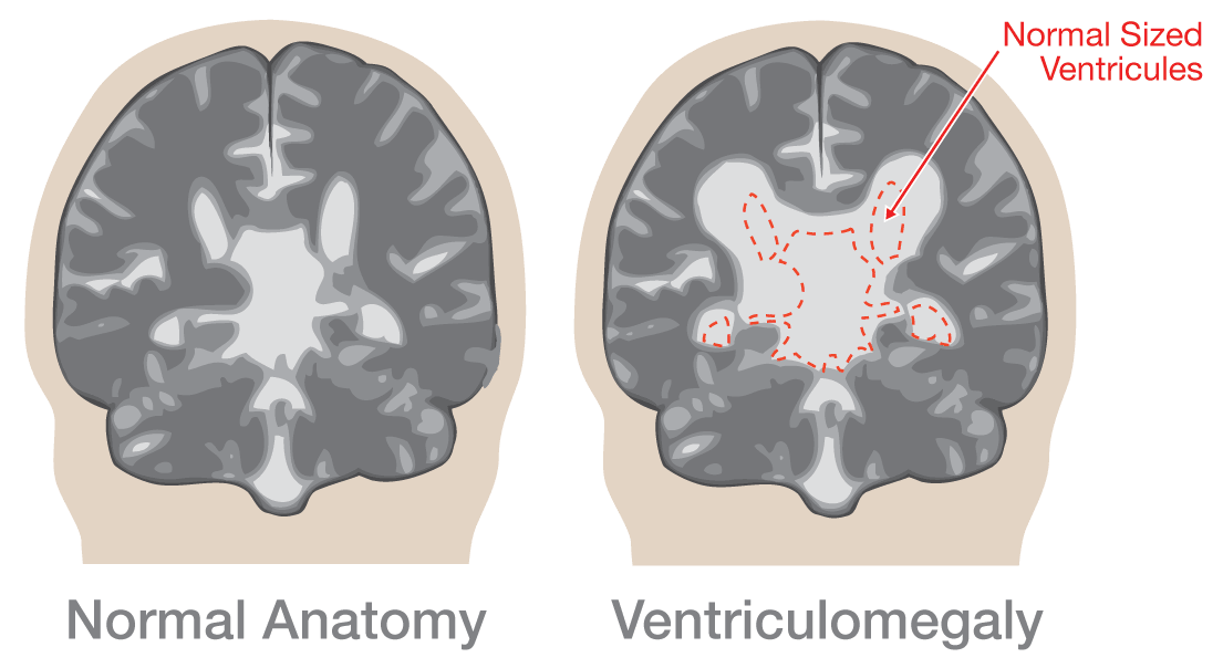
Abnormal accumulation of cerebrospinal fluid within the cerebral ventricles resulting in dilatation of the ventricles. The severity of ventriculomegaly depends on how enlarged the brain is. The ventricular system consists of four ventricles.
Gray bars represent the first occurrence of ventricle enlargement and ensuing time points following EAE. In addition to providing physical protection to the brain tissue it provides the nervous system with nutrients and removes waste among other functions. There are four such hollow spaces in the brain that house cerebrospinal fluid CSF.
It is a biological marker of a past or present brain disorder usually one involving either degeneration of brain tissue or obstruction to cerebrospinal fluid circulation. The causes of enlarged ventricles of brain hydrocephalus are still not well understood. If enlarging ventricles serve as a marker of faulty cerebrospinal fluid CSF clearance mechanisms then a relationship may be demonstrable between increasing ventricular volume and decreasing levels of amyloid beta peptide Aβ in CSF in preclinical and early AD.
2006 non-DS mental retardation Spencer et al. D continue to enlarge for. The choroid plexuses are located in the ventricles produce.
As a result the pressure within the skull increases and the ventricles enlarge. C are most likely due to medication. Cerebral ventricular enlargement is a non-specific alteration in gross brain structure the end result of numerous pathological processes and even more numerous etiologies.
Hydrocephalus may result from inherited genetic abnormalities such as the genetic defect that causes aqueductal stenosis or developmental disorders such as those associated with neural tube defects including spina bifida and encephalocele. Ventriculomegaly is commonly observed in most neurodegenerative disorders and results from passive enlargement of the lateral third and fourth ventricles following brain parenchymal shrinkage. Healthy siblings share third ventricle enlargement with their affected relatives and may partially display a reduction in cerebral volume.
The meninges of the brain and spinal cord are continuous being linked through the magnum foramen. In some cases fluid keeps building up and the ventricles grow larger. The brain ventricular system is in charge of cerebrospinal fluid CSF production which is essential for its normal function.
Its purpose is to help protect the central nervous system and supply it with nutrients. Cerebrospinal fluid or CSF is made and stored in the brains ventricles. Create your own Quiz.
Enlarged ventricles are due to general brain atrophy an age related condition especially recognizing that he has no neurological deficits. Our data suggest that changes in ventricle size during the early stages of brain inflammation could be an early indicator of the events preceding neurological disease and warrant further exploration in preclinical and clinical studies. This article will look at the structure of this system and how it helps the brain.
2005 fragile X mental retardation Reiss et al. Ventriculomegaly is a condition in which the brain ventricles or fluid-filled cavities are enlarged due to build up of cerebrospinal fluid CSF. Nevertheless ventriculomegaly has a large effect size in AD and is a.
Only the group of 60 to 80 year old patient. B are seen in childhood but have typically disappeared by adulthood. Enlargement of the lateral ventricles following TAI and TAI with hypoxia TAIHx was quantified by expressing the ventricle size as percentage of the.
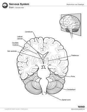
Two lateral ventricles a third ventricle and a fourth ventricle. What do enlarged brain ventricles suggest in. MRI scans indicate that persons with schizophrenia tend to have enlarged ventricles which suggest that the surrounding brain tissue has shrunk.
There are four specialized brain ventricles composed of the following. In 56 patients there was no correlation between the CT findings of enlarged ventricles and sulci and clinical findings of psychoorganic syndromes. Patients with MS exhibited ubiquitous ventricle enlargement whereas NMOSD ventricle enlargement is mainly in the lateral and 3rd ventricles.
Asked Apr 11 2017 in Psychology by mi_flux. Commonly associated with additional fetal anomalies. These findings suggest that.
Infants with Down syndrome trisomy 21 have been shown to have significant enlargement of the third ventricle in both width and length despite the smaller overall head circumference. The ventricles of the brain are a communicating network of cavities filled with cerebrospinal fluid CSF and located within the brain parenchyma. 2000 Klinefelter syndrome a sex chromosome aneuploidy Itti et al.
The fluid cerebrospinal fluid is produced in the ventricular system of the brain. Asked Apr 6 2016 in Psychology by Platini. The ventricular system is composed of 2 lateral ventricles the third ventricle the cerebral aqueduct and the fourth ventricle see the images below.
The Cerebral Cortex is made up of tightly packed neurons and is the wrinkly outermost layer that surrounds the brain and is divided into lobes. Significant ventricular enlargement has been associated with AD 12 15 but it is a rather nonspecific finding. The enlargement of brain ventricles has been described for various brain disorders with cognitive impairment including multiple sclerosis Bakshi et al.
Enlarged brain ventricles seen in people with schizophrenia. Which of the following is a computer-enhanced three-dimensional representation of the brain that responds to the bodys magnetic field. Obstructive communicating or posthemorrhagic hydrocephalus would be.
Genetic malformations such as congenital aqueductal stenosis can cause enlargement of the third ventricle. Compression of developing brain tissue and brain damage may result. It happens when one or more ventricals which are normally hollow areas in the brain have too much cerebrospinal fluid.
Hydrocephalus can be present at birth due to a genetic or developmental abnormality. CSF is a fluid that protects the brain and spinal cord. Intracranial volume CSF volume or volumes of the cerebellum amygdala hippocampus or the parahippocampal gyrus did not significantly differ among the patients siblings and comparison subjects.
Different patterns of ventricle enlargement in MS and NMOSD. Hydrocephalus is a life-threatening medical condition in which cerebrospinal fluid gets blocked and builds up in the ventricles or subarachnoid space. Test your understanding by taking this neuro cortex meninges ventricles MCQs.
The other choices are not correct because of age congenital hydrocephalus is seen in the very young and the lack of lesions in the brain or ventricles.

Quantification Of The Asymmetry Between Right And Left Cerebral Lateral Ventricles By Indexing Methods

Pin Van Nonas Arc Op Ivemark Syndrome

Cerebral Ventricle An Overview Sciencedirect Topics

Pdf Cerebral Lateral Ventricular Asymmetry On Ct How Much Asymmetry Is Representing Pathology

Mri Of The Head Showing Asymmetrical Enlargement Of The Right Lateral Download Scientific Diagram

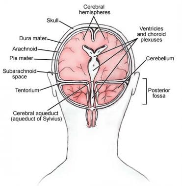
Posterior Fossa

Pin On Endocrine System

Fetal Ventriculomegaly Lurie Children S
Cerebrospinal Fluid Dynamics Relevant To Hydrocephalus

Pin By Menasia On Anatomy Nurse Radiology Medical
Learn About A Ventriculomegaly Diagnosis During Pregnancy
Ventricles Of The Brain Overview Gross Anatomy Microscopic Anatomy

References In Perioperative Management Of Hydrocephalus Bja Education

Management Of Post Hemorrhagic Ventricular Dilatation In The Infant Born Preterm The Journal Of Pediatrics
3d Heart Ems Pinterest Heart And 3d
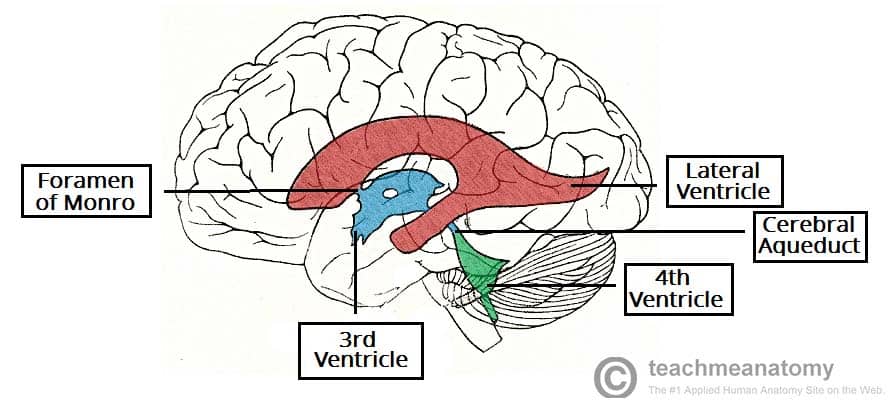
The Ventricles Of The Brain Lateral Third Fourth Teachmeanatomy
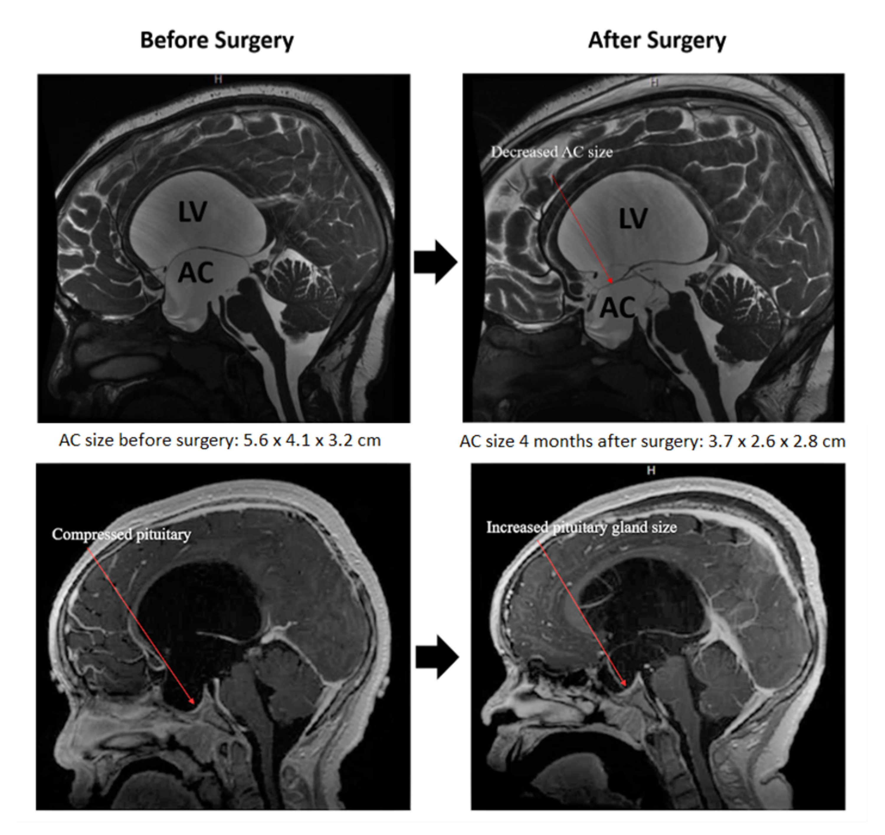
Cureus Syndrome Of Inappropriate Antidiuretic Hormone Secretion Siadh And Precocious Puberty With A Third Ventricle Arachnoid Cyst
Ventricles Of The Brain Overview Gross Anatomy Microscopic Anatomy

The Four Csf Filled Ventricles In The Adult Mouse Brain Highlighted In Download Scientific Diagram


Comments
Post a Comment